
In managing hip fractures it is important that you understand some basic concepts that underpin the decision making.
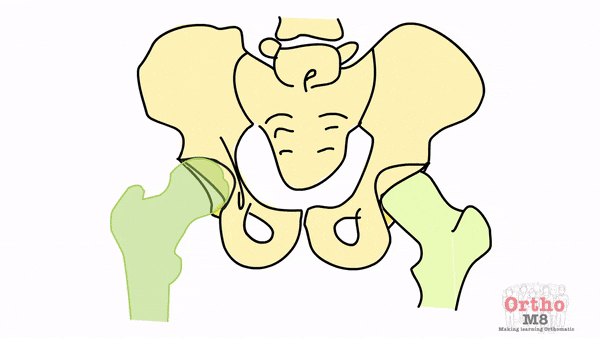
In orthopaedics what we mean by the hip is the proximal femur and its articulation with the acetabulum.

Embryology
The hip develops from birth through to adulthood therefore being aware of the stage of development helps decision making. For example a hip fracture in a skeletally immature patient will be treated differently to that of a young adult and even an elderly patient.

The proximal femur can be divided into the following parts
- Head
- Neck
- Greater trochanter
- Lesser trochanter
- Subtrochanteric region

The proximal femur typically has certain parameters that we have identified in the general population for the neck shaft angle and the degree of anteversion of the neck. It is important to understand the normal variants in order to identify pathology.
The normal neck shaft angle is 130+/-7 degrees and the anteversion is 10+/-7 degrees.
The Blood supply of the proximal femur is made up of the branches of the medial and lateral femoral circumflex arteries as well as the artery of the ligamentum teres. It is retrograde in nature, starting distally and coursing proximally to supply the femoral head. This is generally unusual but also occurs in other bones that we take great care to identify injuries to such as the Scaphoid, Talus and Proximal humerus to name a few.
There is a capsule that covers the head and neck as well as strong ligaments that help to stabilise the hip.
There are strong muscles that attach to the hip and act as dynamic stabilisers as well as enabling locomotion. These must be known for surgical approaches and should be considered in treatment decisions.